Knee Muscle Anatomy Mri : Paediatric Knee Mri Indications For Gp Referred Medicare - From superficial to deep includes the pes anserinus tendons, semimembranosus tendon, tibial collateral ligament, meniscofemoral and meniscotibial ligaments, and the medial meniscus.
Knee Muscle Anatomy Mri : Paediatric Knee Mri Indications For Gp Referred Medicare - From superficial to deep includes the pes anserinus tendons, semimembranosus tendon, tibial collateral ligament, meniscofemoral and meniscotibial ligaments, and the medial meniscus.. Anatomy of the knee bones around the knee. Knee muscle anatomy axial mri : Atlas of knee mri anatomy. These muscles work in groups to flex, extend and stabilize the knee joint. The images may also help physicians to distinguish normal, healthy tissues from dead tissues(2).
Doctors may recommend a knee mri if a patient experiences the following(3): This mri knee sagittal cross sectional anatomy tool is absolutely free to use. The hamstrings muscles allow for strength and power in flexion (bending). Intensity corresponds to a pathologic lesion. Mri knee anatomy | knee sagittal anatomy | free cross sectional anatomy.
The Knee Resource On Twitter A Lateral Knee Mri Explained Knee from pbs.twimg.com Knee muscle anatomy axial mri : The images may also help physicians to distinguish normal, healthy tissues from dead tissues(2). Injuries such as anterior cruciate ligament, meniscus and rotator cuff tears are all easily diagnosed when there is a firm understanding and knowledge of human anatomy. Plantaris acts weakly to plantar flex the foot and flex the knee. Magnetic resonance imaging is particularly well suited for the medical evaluation of the musculoskeletal (msk) system including the knee, shoulder, ankle, wrist and elbow. Atlas of knee mri anatomy. Anterior and posterior cruciate ligaments. Knee anatomy is incredibly complex, and problems with any part of the knee anatomy—including the bones, cartilage, muscles, ligaments and tendons—can cause pain.
In this presentation mri anatomy biceps femoris muscle.
Superiorly, it extends to the level of the crossing of the biceps femoris tendon, and remains superficial to fcl in this location.10 Richolt j.a., jakab m., kikinis r. Articular surface of patella and femur, condyle, epicondyle and muscles (popliteus, sartorius, gastrocnemius, semimembranous with tendos.) find this pin and more on anatomyby radiologist.ayman almatboly. From superficial to deep includes the pes anserinus tendons, semimembranosus tendon, tibial collateral ligament, meniscofemoral and meniscotibial ligaments, and the medial meniscus. These muscles work in groups to flex extend and stabilize the knee joint. Doctors may recommend a knee mri if a patient experiences the following(3): C m c j o i n t m c p j o i n t i p j o i n t m e t a c a r p a l p r o x i m a l p h a l a n x jun 17, 2021 · knee joint (articulatio genu) the knee joint is. Louis, usa and the rijnland hospital in leiderdorp, the netherlands. The knee joins the thigh bone (femur) to the shin bone (tibia). Mri anatomy of knee dr. Plantaris can have variable size, but in most cases is difficult to demonstrate on routine mri studies. Related posts of knee muscle anatomy mri anatomy muscle system. To realign the anterior cruciate ligament parallel with the sagittal imaging plane.
The knee joint is a synovial joint which connects the femur thigh bone the longest bone in the body to the tibia shin bone. These muscles work in groups to flex extend and stabilize the knee joint. When a muscle has different orientations of the tendons it means that there are different patterns of edema possible depending on the tendon injured. Knee muscle anatomy axial mri : Coronal anatomy of the knee.
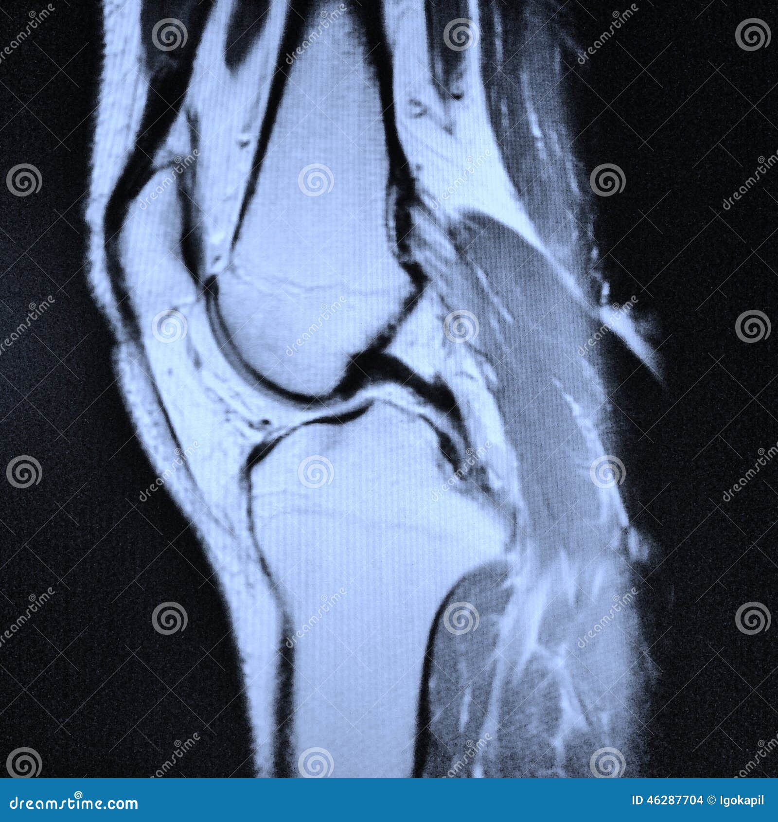
Left Knee Mri Stock Photo Image Of Ligaments Cartilage 46287704 from thumbs.dreamstime.com Prescribe sagittal plane off axial images with line parallel to bony glenoid. Use the mouse scroll wheel to move the images up and down alternatively use the tiny arrows (>>) on both side of the image to move the images.>>) on both side of the image to move the images. Shop your anatomē everyday essentials or add a new product to your collection. Can also generate proton density images. When a muscle has different orientations of the tendons it means that there are different patterns of edema possible depending on the tendon injured. This approach is an example of how to create a radiological report of an mri knee with coverage of the most common anatomical sites of possible pathology. Mri anatomy of knee dr. Knee muscle anatomy axial mri :
An mri of the knee of a healthy subject was performed in the 3 planes of space (coronal, axial, sagittal) commonly used in osteoarticular imaging, with two weightings most commonly used to explore the musculoskeletal pathology of the knee:
Assoc prof craig hacking and dr shu su et al. An mri of the knee of a healthy subject was performed in the 3 planes of space (coronal, axial, sagittal) commonly used in osteoarticular imaging, with two weightings most commonly used to explore the musculoskeletal pathology of the knee: Doctors may recommend a knee mri if a patient experiences the following(3): Mri wrist anatomy scroll using the mouse wheel or the arrows. The knee joint is a complex joint that connects three bones; Knee muscle anatomy axial mri : From superficial to deep includes the pes anserinus tendons, semimembranosus tendon, tibial collateral ligament, meniscofemoral and meniscotibial ligaments, and the medial meniscus. Mri anatomy of knee dr. This approach is an example of how to create a radiological report of an mri knee with coverage of the most common anatomical sites of possible pathology. Anatomy basic knee mri checklist. Coronal anatomy of the knee. Use the mouse scroll wheel to move the images up and down alternatively use the tiny arrows (>>) on both side of the image to move the images.>>) on both side of the image to move the images. Use the mouse scroll wheel to move the images up and down alternatively use the tiny arrows (>>) on both side of the image to move the images.
Knee muscle anatomy axial mri : This approach is an example of how to create a radiological report of an mri knee with coverage of the most common anatomical sites of possible pathology. The images may also help physicians to distinguish normal, healthy tissues from dead tissues(2). Mri knee anatomy scroll using the mouse wheel or the arrows. Assoc prof craig hacking and dr shu su et al.
The Knee Mri Atlas Of Anatomy In Medical Imagery from www.imaios.com Anatomy of the knee bones around the knee. Knee muscle anatomy axial mri : Anatomy basic knee mri checklist. Anterior and posterior cruciate ligaments. Richolt j.a., jakab m., kikinis r. Plantaris can have variable size, but in most cases is difficult to demonstrate on routine mri studies. Can also generate proton density images. Atlas of knee mri anatomy.
Magnetic resonance imaging is particularly well suited for the medical evaluation of the musculoskeletal (msk) system including the knee, shoulder, ankle, wrist and elbow.
C m c j o i n t m c p j o i n t i p j o i n t m e t a c a r p a l p r o x i m a l p h a l a n x jun 17, 2021 · knee joint (articulatio genu) the knee joint is. Plantaris can have variable size, but in most cases is difficult to demonstrate on routine mri studies. The hamstrings muscles allow for strength and power in flexion (bending). Über 80% neue produkte zum festpreis; Abnormal anatomy with normal signal, i.e. From superficial to deep includes the pes anserinus tendons, semimembranosus tendon, tibial collateral ligament, meniscofemoral and meniscotibial ligaments, and the medial meniscus. Assoc prof craig hacking and dr shu su et al. The knee joins the thigh bone (femur) to the shin bone (tibia). Intensity corresponds to a pathologic lesion. Anatomy arthrogram anatomy basic shoulder mri. Related posts of muscle anatomy knee mri muscle anatomy get body smart. Anatomy muscle system 12 photos of the anatomy muscle system anatomy and physiology muscular system exam, anatomy and physiology muscular system labeling quiz, anatomy and physiology muscular system pdf, anatomy and physiology muscular system review, human anatomy muscular system quizzes, human muscles, anatomy and physiology. Related posts of knee muscle anatomy mri anatomy muscle system.


0 Komentar